Monica Moya (17-ERD-054)
Executive Summary
We are investigating how to bio-print and computationally model vascular flow of three-dimensional human tissue structures, which has application to rapid drug discovery, personalized medicine, and therapeutic countermeasures. This research will also support novel applications for three-dimensional bio-printing.
Project Description
A fatal complication in cancer is brain metastasis. The brain microenvironment is of particular interest because only certain cancers (e.g., breast cancer) have an affinity for metastasizing to the brain. Brain metastases are difficult to treat because the neurovascular system is actively involved in protecting metastatic cells during extravasation (i.e., the leakage of intravenously infused, potentially damaging medications into the extravascular tissue around the site of infusion) and proliferation in the brain. We are using three-dimensional bio-printing to create a platform of functioning brain vasculature to provide a modality for engineering human tissues that mimic biological functions. The platform will enable precise control over features and structures to assist with production of accurate predictive computer models. Given the infancy of the field, much of the bio-printing research conducted to date has focused on technology development (print process and materials) with proof-of-concept experiments. We intend to advance existing technology developed at the Laboratory for printing perfused vessels, and apply it to create a human model of brain metastasis to study the dynamic interaction of extravasation that occurs between the cancer cells and the neurovascular system. In addition to serving as an in vitro research platform, this tissue model will also be used to inform and validate a computational fluid-dynamic model that simulates how cancer moves through blood vessels. Combining observations of metastatic breast cancer cells circulating within microscopic vessels printed in three dimensions with advanced computational fluid-dynamic modeling will enable the creation of a testing platform able to predict vascular regions where metastasis is most likely to occur. Our specific goals are to develop new biological inks and methods for creating brain vascular tissue models, create and test informative three-dimensional geometries, and validate our computational flow model. Lastly, we will develop and validate both an experimental and theoretical three-dimensional tissue model of metastatic cancer.
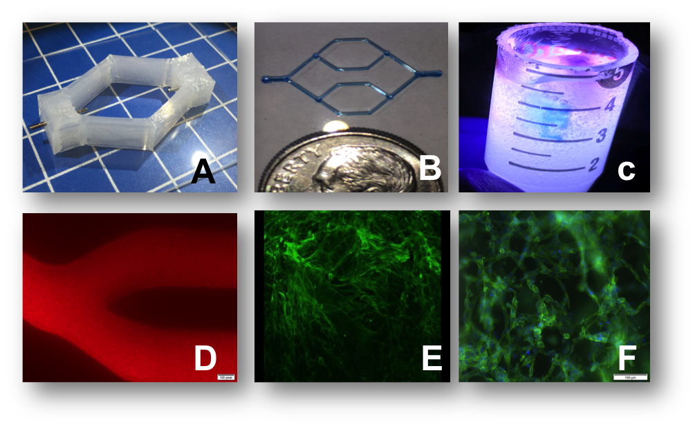
We expect this research to result in important breakthroughs and provide unique insight into the cancer metastatic process. Specifically, we intend to develop a three-dimensional human-tissue model of the blood-brain barrier that will have a range of applications, such as for rapid drug discovery, personalized medicine, and therapeutic countermeasures. In addition, we will produce a biological model capable of predicting the spread of biological infections or agents in complex and dynamic anatomical vascular structures. For this study, we will evaluate the spread of cancer cells, but the modified methodology could have applications for the dynamics of viruses, immune cells, and other biological functions. In addition, using bio-printing to create human-tissue models with dynamic vasculature is key to understanding the spread of infections or agents and, consequently, developing better medical countermeasures against chemical and biological threats. The metastasis computational model we develop could be modified from predicting cancer movement in the vasculature to predicting the spread of non-tumors (i.e., biological and chemical agents). The two technologies we will develop for this effort, bio-printing (see figure) and computer simulations, will contribute to a better understanding of complex biology and enable more precise predictions of health risks.
Mission Relevance
Developing a platform of functioning brain vasculature using the advanced manufacturing capability of three-dimensional printing directly supports the Laboratory's core competency in bioscience and bioengineering, as well as the core competency in advanced materials and manufacturing. Creation of a computational fluid-dynamic model of cell movement through blood vessels is also relevant to the Laboratory's core competency in high-performance computing, computer-based simulation, and data science. This research supports the DOE's goal to deliver scientific discoveries and major scientific tools that transform our understanding of nature and strengthen the connection between advances in fundamental science and technology innovation.
FY17 Accomplishments and Results
A major accomplishment for FY17 was optimizing three-dimensional printing with sacrificial inks that allowed us to create hollow channels within the printed structures. We successfully seeded the channels created by the sacrificial ink with human brain endothelial cells and perfused the cell-seeded vascular channel for ten days under physiological flow conditions. Specifically, we (1) accomplished three-dimensional printing with polydimethylsiloxane (a silicon-based organic polymer) to create bioreactors to house the tissue constructs (previously, three-dimensional printed tissue constructs were cultured in traditionally mold-fabricated bioreactors), which increases our printing capability and allows us to entirely print the biological tissues and the bioreactor housing together in three dimensions; (2) began to develop and optimize new bio-inks specifically for neuronal cells; (3) used one such bio-ink to create a three-dimensional microenvironment with dispersed cells that was successfully cultured for ten days under induced interstitial-like gradients across the cell-laden gel; and (4) began to examine the mechanical microenvironment of printable extracellular matrices.
Publications and Presentations
Moya, M.L. and E. K. Wheeler, 2017. "3D Dynamic Blood Brain Barrier Model." LLNL-PRES-713825.


