Amy Jenei (17-ERD-025)
Executive Summary
To determine whether phase transitions to complex structural forms are ubiquitous at high pressures, we are measuring changes in ionization potential of iron and magnesium under high pressure. These measurements could provide key experimental benchmarking data for theoretical models that could be used in assessing the effectiveness and reliability of the U.S. nuclear weapons stockpile as well as for Inertial Confinement Fusion designs.
Project Description
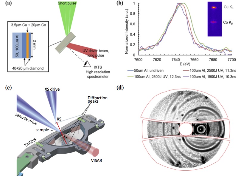
The effect of surrounding electrons on an atom's ionization potential (the energy required to remove an electron from a neutral atom) drives a multitude of fundamental material properties including electron and heat transport, opacity, compressibility, crystal structure, and bonding. When density increases, the enhanced interactions of neighboring atoms increases the energy of bound electrons, which then require less energy to ionize. Equation-of-state (EOS) models for describing stellar and planetary interiors as well as stockpile materials and Inertial Confinement Fusion employ simplified approximations of this changing ionization potential. Recent studies to benchmark these approximations at high pressure and temperature have yielded potentially conflicting results, but each study accesses a different phase-space range, and effects of temperature and density are difficult to decouple. In solid and low-electronvolt temperature regimes, simple approximations for this changing ionization potential (referred to as the ionization potential depression in the plasma phase) are usually inaccurate. We are performing in situ measurements of properties predicted to be sensitive to or correlated with density-driven changes in ionization potential. Specifically, we are examining the absorption edge (a substance's abrupt increase in absorption of electromagnetic radiation as frequency is increased) and the crystal structure to test and benchmark model predictions for ionization potential depression at high density. These predictions will be used to refine EOS models for materials under high pressure and density. Using the OMEGA laser facility at the Laboratory for Laser Energetics, New York, and Livermore's National Ignition Facility (NIF), we are performing x-ray inelastic scattering and x-ray diffraction measurements on iron and magnesium compressed to high density using ramp-shaped laser pulses.
We expect to determine whether the predicted phase transition sequence in iron and magnesium is observed experimentally, to determine the structures of the high-pressure phases, and to investigate the possibility of detecting additional diffraction features from non-nuclear maxima in electron densities (i.e., a maximum electron density that appears at a position away, rather than near, the nucleus) in the material's structural lattice. Experimental data generated will be key for developing ionization models in the high-energy-density regime. We are also experimentally investigating and benchmarking model predictions for high-pressure electride phase formation in magnesium (the formation of an ionic compound in which the axion is an electron). Our specific objectives are to confirm or refute predictions of high-pressure crystal structure and the conditions for transitions, and to find evidence for the presence of non-nuclear maxima in electron densities in the lattice. Successful project results will impact EOS models used for Inertial Confinement Fusion and stockpile stewardship, and will improve understanding of the fundamental processes in Earth’s inner core boundary, such as thermal conduction from the core to the mantle.
Mission Relevance
Our work addresses the NNSA goal of managing and understanding the condition of the nuclear weapons stockpile, and supports the Laboratory’s core competency in high-energy-density science through examination of model predictions for ionization potential depression at high pressure and density.
FY17 Accomplishments and Results
In FY17 we (1) collected high-quality electron-induced fluorescence data for iron on OMEGA, showing non-negligible spectral shift vs. compression; (2) began first-principles molecular-dynamics (FPMD) simulations of electronic structure for iron; (3) collected high-quality data on NIF for magnesium, including results at 300 GPa showing an unexpected new phase; (4) completed FPMD simulations of magnesium over 250–650 GPa and 2500–1000 K; and (5) processed data to extract structural and dynamical properties and charge distributions.


