Tom Pardini (15-LW-002)
Abstract
The goal of this project was to study ultra-fast dynamics in condensed matter using an x-ray pump/x-ray probe experimental set-up for hard X-ray Free Electron Lasers (XFELs). The project consisted of two phases: (1) the design, construction, and calibration of a Mirror-based dElay Line for X-rays (MEL-X) (Pardini et al. 2015); and (2) its deployment at the Linac Coherent Light Source (LCLS, at the SLAC National Accelerator Laboratory) (Bostedt et al. 2016) for a five-day experimental campaign. The successful completion of the project showcased several technical accomplishments related to the construction of a complex delay line for x-rays, such as high throughput, broad energy response, and, most importantly, pointing stability and repeatability. Additionally, it provided new data for the onset of non-thermal melting in single-crystal silicon, a solid-to-liquid phase transition that has been investigated for more than 20 years. The MEL-X, coupled with the LCLS, allowed us to achieve the temporal resolution needed to clearly decouple the electronic and ionic time scales characterizing the melting transition. Our data reveals a never-resolved-before delay in the onset of the transition, which we interpret as the time needed for the electronic system to equilibrate at or above the critical non-thermal melting temperature.
Background and Research Objectives
The advent of XFELs has revolutionized the field of ultra-fast science, by providing the necessary resolution to couple to the characteristic time scales of ultra-fast phenomena. A standard experimental set-up at any XFEL instrument is characterized by a pump beam triggering ultra-fast dynamic, followed by a probe beam capable of characterizing the fast-changing state of the sample. The pump beam is generally provided by an external optical laser, while the x-ray probe beam is provided by the XFEL. There are several limitations to this scheme. On the one hand, the optical pump beam has a much shorter penetration depth than the probe beam, possibly leading to the probing of unperturbed portions of the sample. On the other hand, the timing of the optical-laser pump and the x-ray probe can be susceptible to significant uncertainties. Ultimately, the photon energy of the optical-laser pump is too low to excite core levels in most solids, precluding the study of core-level contribution to ultra-fast transitions. We succeeded in developing an x-ray pump/x-ray probe set-up by splitting a single pulse from an XFEL into two pulses, with a tunable time delay between them. We did so by building a split-and-delay line based on reflective x-ray optics named Mirror-based dElay Line for X-rays (MEL-X). Leveraging Lawrence Livermore National Laboratory’s expertise in x-ray optics, precision engineering, and XFEL science, we were able to design and build a portable delay line that was installed and aligned at an XFEL instrument within the time constraint (approximately two weeks) of a typical experimental campaign. The challenges we faced ranged from (1) developing a tool that could work over a broad range of photon energies, (2) achieving the necessary throughput to trigger the phase transition of interest, (3) guaranteeing stable overlap of the pump and probe beams at the sample plane, and (4) decoupling the system from the many vibrational and thermal instabilities present at an XFEL beamline.
Once deployed at the LCLS, we used the MEL-X to study the effect of core-level excitations (via x-ray pumping) on the time scale of ultra-fast melting of silicon. This effect has been experimentally observed in silicon (Sokolowski-Tinten et al. 1995), gallium arsenide (Rousse 2001), indium antimonide (InSb) (Lindenberg et al. 2005; Gaffney et al. 2005; Hillyard et al. 2007), and germanium (Siders et al. 1999) triggered via optical lasers. It is well understood (Recoules et al. 2006; Hillyard et al. 2008, Silvestrelli et al. 1996; Stampfli et al. 1995; Shank et al. 1983) that ultra-fast melting is initiated by the photo-excitation of a large number of electrons into the conduction band. Such excitation begins with photo-absorption by a few primary electrons, followed by a cascading effect that leads to secondary electron generation. Above some threshold electronic temperature, a significant number of bonding states are depleted and anti-bonding states are populated. At this stage, ions experience a significant change in the electrostatic potential, leading to inertial displacement from their equilibrium positions. Recoules et al (2006) found that, for Si, a threshold electronic temperature of 1.5 eV/atom is enough to cause an ultra-fast lattice instability. The latter is named non-thermal melting and is caused by the lack of thermal equilibration between the electronic and ionic systems.
All experiments to date have used an optical laser pump to trigger the transition, coupling directly to valence-band electrons. These electrons have low kinetic energies and thermalize quickly, leading to a fast population of antibonding states. By accessing core-level electrons with our x-ray pump, we set out to quantify the change in the transition time scale caused by the large kinetic energy of the photo-electrons after excitation. Recent calculations have quantified the time scale of thermalization of fast photo-electrons originating from core levels. Such a thermalization time scale is longer than the equivalent time scale for valence electrons, given that it is proportional to the number of electron-electron collisions, which scales with energy. The successful first experimental campaign conducted with the MEL-X enabled us to quantify the ultra-fast melting time scale in the x-ray pump regime.
Scientific Approach and Accomplishments
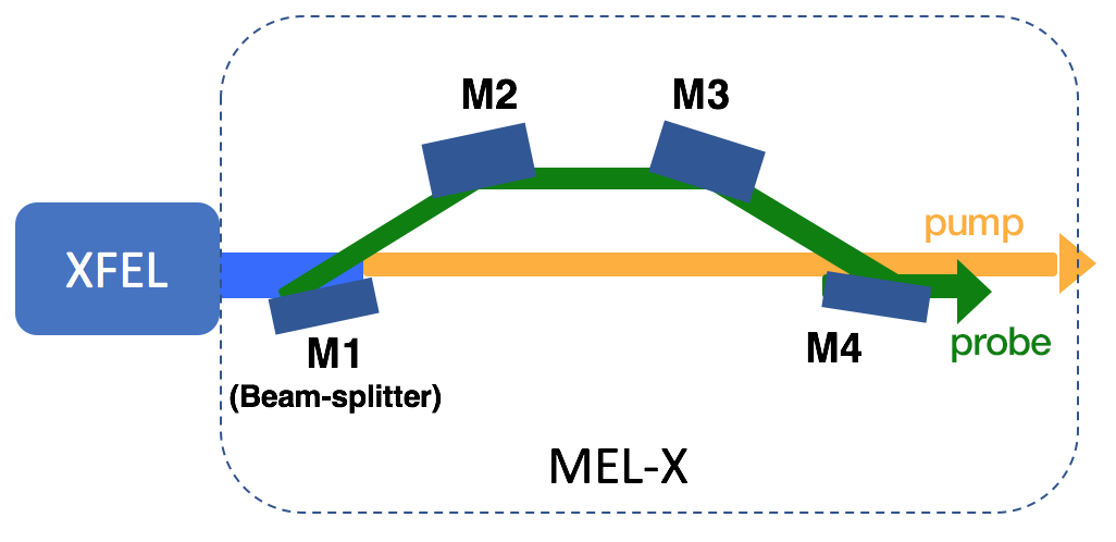
A schematic of the MEL-X is shown in Figure 1.
The first mirror (M1) acts as a beam-splitter, separating the incoming LCLS beam in a pump beam and a probe beam. The pump beam proceeds to the focal plane with no reflections, retaining its full intensity. The probe beam undergoes four reflections before reaching the focal plane. The relative position of mirrors 2 and 3 (M2, M3) determines the path length difference between the pump and the probe, and therefore the delay time. Mirror 4 (M4) recombines the two beams at the focal plane.
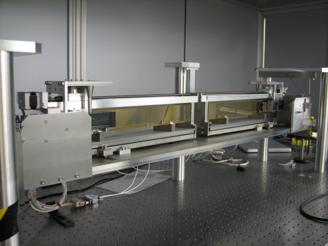
The assembled MEL-X used during the calibration phase at Lawrence Livermore National Laboratory is shown in Figure 2.
Figure 2. The Mirror-based dElay Line for X-rays (MEL-X) setup used during the alignment phase at Lawrence Livermore National Laboratory.
The main set of the MEL-X's specifications is listed in Table 1.
To achieve the necessary throughput to trigger ultra-fast melting, each MEL-X optic was coated with 300 Å of iridium, providing nearly 79 percent reflectivity (for each reflection) up to 8 keV. The coatings were developed and characterized by our group at Livermore, and demonstrated the feasibility of metallic coatings for high-fluence applications. In fact, the damage that was believed to take place in high-Z-axis coatings at high fluence is reduced to negligible amounts by the large amount of energy released by photoelectrons. The absence of surface damage on the MEL-X optics confirmed this concept. Additionally, a metallic coating working below its critical angle provides almost flat response up to 8 keV photons. This was an important requirement for the MEL-X, as it was unclear what photon energy was going to be best suited for the first experimental campaign.
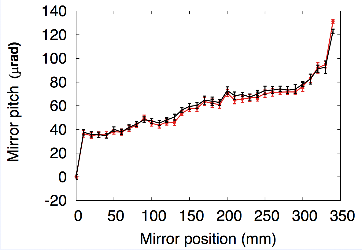
The pointing precision and repeatability of the MEL-X were key deliverables of this project. In fact, the time constraint of an XFEL experiment would not allow a user to spend much time aligning the pump and probe beam at the sample plane. We achieved such precision by designing a highly accurate transport mechanism for the M2 and M3 mirrors, responsible for setting the delay time. To avoid alignment instability caused by wobble in commercially available motors, the MEL-X features a custom-built translation stage. Both M2 and M3 are mounted on carriages that physically slide on top of polished Zerodur rails. These rails have been polished to a λ/10 specification for figure error (λ = 633 nm). The coupling between the carriages and the rails is provided by custom-made polyamide pads, chosen for the negligible friction in vacuum. Figure 3 shows the pitch of the M2 mirror measured with a digital auto-collimator as a function of M2 position, over two separate M2 scans. Similar data was taken on the M3 mirror. The highly reproducible pitch values translate in highly reproducible positioning of the probe beam at the sample plane.
The MEL-X is suspended from the lid of its vacuum chamber by six stainless-steel rods. This is done to decouple the MEL-X as much as possible from vibrational and thermal instabilities typical of a beamline setting.
On November 24, 2016, the MEL-X began its first five-day experimental campaign at the Coherent X-ray Imaging (CXI) instrument (Liang et al. 2015) at LCLS. The goal of the experiment was to measure the disappearance of Bragg signal from silicon as a function of the time delay between pump and probe beams. In fact, the disappearance of Bragg signal is an indication of a loss of crystal order, which implies a disorder transition. To this end, the MEL-X was installed four meters upstream of the sample plane, and approximately four meters downstream of the focusing Kirkpatrick-Baez (KB) optics. Figure 4 is a schematic diagram of the experimental set-up.
The XFEL was operated in self-amplified spontaneous emission (SASE) mode, with an average pulse energy of 2.43 mJ, and a photon energy of 5.95 keV. The length of the pump and probe pulses was 25 fs, and the uncertainty on their delay was ± 40 fs. The latter was caused by an intrinsic uncertainty on the initial position of the M2 and M3 mirrors. A spectrometer deployed in parasitic mode measured the spectral content of each x-ray pulse. The silicon (333) Bragg reflection was measured on a pixel array detector in near back-scattering geometry. A 200 µm-thick silicon single-crystal attenuator with variable transmission was placed between the detector and the sample to limit the number of photons to within the dynamic range of the detector. The detector output was normalized by the spectrometer output after proper calibration. The sample consisted of silicon pillars etched in a 1mm-thick silicon (111) wafer. Details on its engineering are discussed in Pardini et al (2014). We collected data at four delay times: 150 fs, 220 fs, 315 fs, and 385 fs. For each delay investigated, data was collected in two modes: with pump and probe beams separated by 50 µm at the focal plane, and with pump and probe beams overlapped. In both modes, the integrated signal from pump and probe was simultaneously measured on the detector. For the non-overlapped mode, given that the beams hit different spots on the sample, the integrated detector signal is proportional to the unperturbed silicon single-crystal reflectivity, independent of delay time. On the contrary, for the overlapped mode, the integrated detector signal enables extraction of the probe reflectivity as a function of delay once the relative intensity of the pump and probe beams is known.
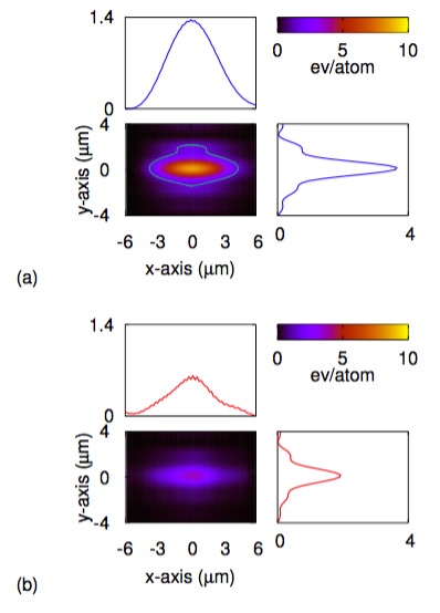
The x-ray pump/x-ray probe nature of this experiment required a detailed knowledge of the pump and probe beam-intensity profiles. To this end, we leveraged our group’s expertise in numerical simulations of coherent beams and damage effects from high-fluence x-ray pulses. Post-mortem analysis of the samples, combined with detailed wavefront-propagation simulations allowed us to determine to a high degree of accuracy the intensity profile for both pump and probe beams at the sample plane. The results of our numerical simulations are shown in Figure 5.
Figure 5. Simulated beam intensity profile at the focal plane for (a) pump and (b) probe beam. The contour line in (a) indicates the threshold fluence of 1.5 eV/atom.
We conclude that the pump and probe beams generated by combining the MEL-X with the LCLS were elongated along the x-axis, as expected from edge diffraction broadening off the MEL-X beam splitter mirror. Also, we quantified the beam size to be approximately 5 µm x 1.9 µm full width at half maximum along the x-axis and y-axis for both beams.
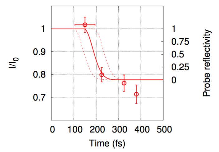
Figure 6 shows the main result of our experimental campaign.
The signal measured on the detector is plotted as a function of pump-probe delay. The right y-axis shows the corresponding probe reflectivity. Temporal error bars are shown for the t=0 data point only. In fact, while the initial position uncertainty on two of the MEL-X mirrors caused an absolute ± 40 fs uncertainty, the relative uncertainty among different delay times is negligible given that the two mirrors are moved with micrometer precision. From the data, we draw the following conclusions. No sign of atomic motion is detected within 150 ± 40 fs from photo-absorption. At 220 fs, the probe Bragg signal has dropped to approximately 0.12 percent. Finally, after 315 fs no probe Bragg reflectivity is observed, indicating a complete loss of long range atomic order. The solid red line is the loss in reflectivity one would expect in the case inertial dynamic, as proposed by Lindenberg et al (2005). According to this model, the loss of crystal order is caused by the sudden lack of restoring force keeping the atoms together, leading them to escape their equilibrium position with a velocity consistent with room-temperature energies. Our data agree with this model. The curve has been fitted to the data with the only degree of freedom being the temporal onset of the transition, which we find to be at 150 fs. Two dashed lines rigidly bound this curve according to the absolute temporal uncertainty of ± 40 fs. Ultimately, we conclude that, for silicon pumped with hard x-rays, the onset of non-thermal melting must happen for t > 110 fs. For the first time, these results provide a direct, quantitative picture of non-thermal melting in a semiconductor where the electronic contribution is resolved from the ionic contribution. The actual time scale of the electronic contribution is a considerable portion of the overall transition, and can be understood as follows. Given the pump dose imparted to the sample, we conclude that about 1 out of 1,000 atoms participate in photoabsorption. Therefore, approximately 0.1 percent of the atoms will have a deep core-hole left behind, which is quickly filled during Auger production, leading to the formation of a valence hole within 10 fs from excitations (Medvedev et al. 2013). At this stage, no appreciable change in electrostatic potential has occurred, since only 0.1 percent of valence electrons have been perturbed, and most bonding states are still occupied. The absorbed pulse energy is stored in the high kinetic energy of the photoelectrons that begin down-scattering with neighboring valence electrons. This initiates the cascading effect, which excites more valence electrons into the conduction band. Medvedev et al (2013) showed that, in SiO2 excited by an 8 keV pulse, the maximum electron density in the conduction band is reached approximately 60 fs after photo-excitation, as each primary electron must undergo over 400 collisions. In the context of our experiment, 60 fs represents a lower bound estimate for the electronic equilibration time as the calculations of Medvedev et al (2013) were done for maximum values of electron densities in the conduction band in the 1018 e-/cm3 range. This value is about four orders of magnitude smaller than what we achieved in our experiment. Our data for the onset of the non-thermal melting transition is consistent with this picture, and indicates that for silicon at least 110 fs are needed to equilibrate the electronic system above the threshold temperature of 1.5 eV/atom.
Impact on Mission
A multilayer delay line for x-ray technology will benefit experiments in ultrafast transitions to warm dense matter, which are relevant to material behavior under extreme conditions, the understanding of planetary formation and composition, and high-energy-density science in general. This research, therefore, aligns well with Lawrence Livermore National Laboratory's core competencies in advanced materials and manufacturing, and high-energy-density science.
This project has further established the Laboratory's expertise in the science and technology of ultra-fast dynamics studied via XFELs, and a follow-up project has been endorsed to develop a new delay line that would reach delays up to the nanosecond scale, enabling the study of phase transition in materials of interest to the Laboratory.
This project has given us the opportunity to strengthen our ties with the LCLS staff, which was instrumental in the success of the experimental campaign. This collaborative experience has led to a proposal by the CXI team to install the MEL-X permanently at the CXI instrument.
Finally, the expertise acquired during this project has motivated Los Alamos National Laboratory to seek a collaboration with our group to work on pump and probe experiments at the Argonne National Laboratory Advanced Photon Source. A preliminary experimental campaign was conducted at APS at the end of the third year of this project. It is expected that this collaboration will continue in the future, possibly merging with programmatic activities of both national laboratories.
Conclusions
Our work enabled x-ray pump/x-ray probe experiments at XFELs by building and deploying a custom split-and-delay line at the LCLS. We demonstrated high throughput, broad spectral response, and extremely stable operations by leveraging Livermore's expertise in x-ray optics, precision engineering, and XFEL science. We have used the MEL-X to study non-thermal melting in single-crystal silicon pumped with 5.95 keV x-rays pulses. The temporal resolution of our experimental set-up allowed us to decouple the time scale for the electronic and ionic contribution to the transition. Our data indicates that, for silicon pumped with hard x-rays, at least 110 fs are needed for the electrons to thermalize at or above the threshold temperature of 1.5 eV/atom. We propose that this rather long time scale is caused by several hundred collisions necessary to thermalize the fast photoelectrons.References
Pardini, T., et al. 2015. “The MEL-X Project at the Lawrence Livermore National Laboratory: A Mirror-Based Delay Line for X-rays.” Proceedings of the SPIE 9589. doi: 10.1117/12.2186796.
Bostedt, C., et al. 2016. "Linac Coherent Light Source: The First Five Years." Reviews of Modern Physics 88. doi: 10.1103/RevModPhys.88.015007.
Gaffney, K., et al. 2005. "Observation of Structural Anisotropy and the Onset of Liquidlike Motion During the Nonthermal Melting of InSb." Physical Review Letters 95.
Hillyard, P. B., et al., 2007. "Carrier-Density-Dependent Lattice Stability in InSb." Physical Review Letters 98. doi: 10.1103/PhysRevLett.98.125501.
——— 2008. "Carrier-Induced Disordering Dynamics in InSb Studied with Density Functional Perturbation Theory." Physical Review B 77 (19): 195213. doi: 10.1103/PhysRevB.77.195213.
Liang, M., et al. 2015. “The Coherent X-ray Imaging instrument at the Linac Coherent Light Source.” Journal of Synchrotron Radiation 22, 514—519. doi: 10.1107/S160057751500449X.
Lindenberg, A., et al. 2005. "Atomic-Scale Visualization of Inertial Dynamics." Science 308, 392—395. doi: 10.1126/science.1107996.
Medvedev, N., et al. 2013. "Electron Kinetics in Femtosecond X-Ray Irradiated SiO2." Contributions to Plasma Physics 53 (4-5): 347—354.
Pardini, T., et al. 2014. "Silicon Mirrors for High-Intensity X-Ray Pump and Probe Experiments." Physical Review Applied 1. doi: 10.1103/PhysRevApplied.1.044007.
Recoules, V., et al. 2006. "Effect of Intense Laser Irradiation on the Lattice Stability of Semiconductors and Metals." Physical Review Letters 96 (055503). doi: 10.1103/PhysRevLett.96.055503.
Rousse, A., et al. 2001. "Non-Thermal Melting in Semiconductors Measured at Femtosecond Resolution." Nature 410: 65—68. doi: 10.1038/35065045.
Shank, C. V., et al. 1983. "Femtosecond-Time-Resolved Surface Structural Dynamics of Optically-Excited Silicon." Physical Review Letters 51, 900.
Siders, C., et al. 1999. "Detection of Nonthermal Melting by Ultrafast X-ray Diffraction." Science 286 (5443): 1340—1342. doi: 10.1126/science.286.5443.1340.
Silvestrelli, P., et al. 1996. "Ab Initio Molecular Dynamics Simulation of Laser Melting of Silicon." Physical Review Letters 77 (15): 3149. doi: 10.1103/PhysRevLett.77.3149.
Sokolowski-Tinten, K., et al. 1995. “Ultrafast Laser-Induced Order-Disorder Transitions in Semiconductors.” Physics Review B51, 14186.
Stampfli, P., and K. Bennemann. 1995. "Theory for the Laser-Induced Femtosecond Phase-Transition of Silicon and GAAS." Applied Physics A 60 (2): 191—196.
Publications and Presentations
Pardini, T., et al. 2015. “The MEL-X project at LLNL: A Delay Line for X-ray Pump/X-ray Probe Experiments at the LCLS." International Workshop on X-ray Mirrors, Lawrence Berkeley National Laboratory, Berkeley, CA, 2015. LLNL-PRES-674343-DRAFT.
——— 2016. “First Experimental Results Obtained with the MEL-X Deployed at the LCLS.” Photon Diagnostic Workshop, SLAC National Accelerator Laboratory, Stanford, CA, 2016. LLNL-PRES-730122-DRAFT.


